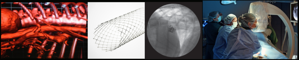
Procedure
This is a minimally invasive, catheter-based procedure performed under anesthesia. Using flouroscopy, a stent is placed in the vena cava via the jugular vein. Embolization coils are then placed in the shunt to reduce blood flow.
Why Take this Procedure
This can reduce flow through a single congenital intrahepatic portosystemic shunt and improve liver function.
Patient Eligibility
Dogs should be more than 5 months old (more than 8 months if a giant breed) and be medically managed for at least 4 weeks (lactulose, antimicrobials, proton pump inhibitor, low protein diet). Anticonvulsant therapy (levetiracetam) should be administered for at least a week prior to shunt attenuation. Advanced imaging under anesthesia (CT angiography) is needed before stent and coil placement to confirm shunt type and location.
Cost
$9000-$11,000 for both CT imaging (to determine the anatomy of the shunt and measure the vena cava) and stent and coil placement.
Length of Stay
1-2 days for initial evaluation and CT study. 2-3 days for stent and coil placement.
Potential Complications
Major complications are uncommon but include portal hypertension, seizures, gastric bleeding, and displacement of the stent or coils.
Anticipated Outcome
We expect to see improvement in hepatic function and mitigation of clinical signs within the first few weeks. In some cases, additional coils are placed after 1-2 months to address continued flow through the shunt.
Contact
If you have any questions, please contact the Texas A&M Interventional Radiology & Endoscopy Service via email at guidewire@cvm.tamu.edu or by phone at 979-845-2351.