What is cranial cruciate ligament rupture?
The cranial cruciate ligament (CrCL) is a fibrous band of tissue located deep within the stifle joint (knee). This ligament is responsible for maintaining joint stability when your pet is standing, walking, or running. Tearing or “rupture” of the CrCL causes joint instability, which leads to joint swelling, pain, and lameness. In dogs, osteoarthritis (degenerative joint disease) will develop if this instability is not corrected. Rupture of the CrCL is one of the most common causes of hind limb lameness in dogs, and it occurs less commonly in cats.
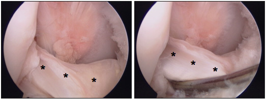
Arthroscopic view of a normal cranial cruciate ligament (asterisks) in the right knee. Note the smooth, taut appearance of the ligament beneath the probe in the right panel.
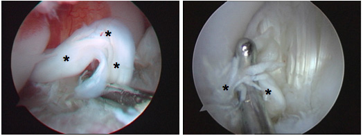
Two examples of ruptured cranial cruciate ligaments from right knees (asterisks). In the left panel the ligament is loose (incompetent) and non-functional. The ligament is being examined with an arthroscopic probe. In the right panel the ligament is completely ruptured and the ligament fibers are frayed and rounded. A probe is positioning the ruptured ligament for photography.
Humans have a similar ligament in their knee, the anterior cruciate ligament (ACL). Most humans tear their ACL during certain athletic maneuvers. Dogs rupture the CrCL either acutely (suddenly) or chronically (over time). Dogs with an acute rupture show no evidence of hind limb lameness or pain before the injury. They suddenly develop severe hind limb lameness, usually during strenuous activity. In contrast, dogs with a chronic rupture develop slow, progressive, hind limb lameness and reluctance to exercise that comes and goes over weeks to months.
Once the CrCL is ruptured, there is an increased risk for damaging other support structures within the knee. One important supporting structure that is commonly damaged along with CrCL rupture is the medial meniscus, a C-shaped piece of fibrocartilage that functions as a shock-absorber and allows the bones above and below the knee joint to interact more effectively.
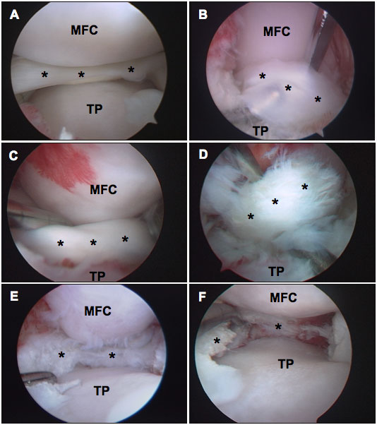
LEGEND: * (asterisk): meniscus | MFC: articular cartilage of the femur | TP: articular cartilage of the tibia
Arthroscopic view of the medial meniscus. A) Normal, healthy meniscus. B,C,D) Representative examples of the classic “bucket handle tear” of the medial meniscus. These tears are detected by examining the meniscus with an arthroscopic probe. Once detected, the injured portion of the meniscus is removed, which eliminates pain and improves limb use after surgery. E,F) Examples of the remaining medial meniscus after removal of meniscal tears.
What are the treatment options for cranial cruciate ligament rupture?
We recommend surgical stabilization of the knee in almost all cases of CrCL rupture. As in humans, many procedures have been developed to stabilize the affected knee. None of these procedures is perfect, meaning that there is not 100% success in every case. The two common procedures we perform at Texas A&M are the tibial plateau leveling osteotomy (TPLO) and the extracapsular stabilization (“Extra-cap”). Depending on your pet’s age, size, and activity, we may recommend one procedure over the other. In many cases, your pet may qualify for either procedure. Regardless of which procedure is performed, we typically begin the surgery by examining the knee joint using arthroscopy. During this part of the surgery, the inner structures of the joint, such as the torn ligament and meniscus, are examined and treated using small skin incisions, an arthroscopic camera, and small arthroscopic instruments. Advantages of arthroscopic treatment include smaller skin incisions, decreased blood-loss during surgery, improved visualization and treatment of the internal structures of the joint, less pain after surgery, and a faster return to using the leg.
Occasionally dogs or cats with CrCL rupture will not qualify for surgery, most commonly because they are at high risk for anesthesia or have other life-threatening medical conditions. In these cases we recommend treating your pet’s knee pain and arthritis with conservative therapy. Conservative treatments may include administration of joint supplements (Adequan ®, Cosequin ®, Dasuquin ®), pain medications, weight loss, rehabilitation, and in some cases administering injections of anti-inflammatories and joint lubricants directly into the joint. Many dogs can be made comfortable for some time with conservative treatment; however, arthritis, pain, and lameness eventually worsen.
If my pet has ruptured its cruciate ligament and I want to pursue additional consultation or treatment at Texas A&M, how can I schedule an appointment?
Appointments can be scheduled with the Orthopedic Surgery Service by contacting the Veterinary Medical Teaching Hospital Monday through Friday at 979-845-2351. Either you or your veterinarian can make the initial phone call, but we will need to speak with your veterinarian prior to confirming the final appointment.