- Chronic Degenerative Valve Disease (CVD)
- How does the normal heart work?
- What is Chronic Degenerative Valve Disease?
- Who develops chronic degenerative valve disease?
- General risk factors for the development of CVD.
- What causes CVD?
- What happens to my dog when it gets CVD?
- Clinical signs (clues) that your dog may have ‘symptomatic’ CVD and needs to be examined by your veterinarian.
- Clinical signs that can be associated with CVD.
- How can my veterinarian determine if my dog has CVD?
- What tests might be recommended by my veterinarian once my dog has CVD?
- Asymptomatic dogs with CVD:
- Symptomatic dogs with CVD:
- Possible tests for dogs with asymptomatic and symptomatic CVD:
- How is CVD treated in dogs?
- Asymptomatic CVD:
- Can I slow down or reverse the progression of CVD in my dog?
- Symptomatic CVD (heart failure):
- Common medications used to treat heart failure due to CVD:
- Common side-effects of medication used to treat heart failure due to CVD:
- What kind of follow-up will my dog need now that it has CVD?
- Recommended follow-up in dogs with asymptomatic CVD (Stage B1 and B2):
- Recommended follow-up in dogs with symptomatic CVD (Stage C):
- Can special diets help dogs with CVD live longer?
- Are there any dietary supplements that may help dogs with CVD live longer?
- What about exercise in my dog once it has CVD?
- Is there a surgical treatment option for dogs with CVD?
- How long will my dog live now that they have CVD?
- Asymptomatic CVD (Stage B1 and B2):
- Symptomatic CVD (Stage C or heart failure):
- Dilated Cardiomyopathy (DCM)
- How does the normal heart work?
- What is dilated cardiomyopathy (DCM)?
- What types of dogs does DCM affect?
- General risk factors for DCM:
- What are the causes of DCM?
- What happens to my dog when it gets DCM?
- Clinical signs that may be associated with DCM in dogs, include:
- How can my veterinarian determine if my dog has DCM?
- Screening tests for DCM that my veterinarian may recommend:
- What tests might be recommended by my veterinarian once my dog has DCM?
- Asymptomatic dogs with DCM:
- Symptomatic dogs with DCM (heart failure and or abnormal heartbeats):
- How is DCM treated in dogs?
- Asymptomatic dogs with DCM:
- Symptomatic dogs with DCM?
- Common medications used to treat heart failure and abnormal heart beats due to DCM:
- Common side-effects of medication used to treat heart failure and or abnormal heart beats due to DCM, include:
- What kind of follow-up will my dog need now that it has DCM?
- Recommended follow-up in dogs with asymptomatic DCM (Stage B):
- Recommended follow-up in dogs with symptomatic DCM (heart failure and or abnormal heartbeats):
- Why should I evaluate my dog’s resting/sleeping breathing rate at home?
- Can special diets help dogs with DCM live longer?
- Are there any dietary supplements that may help dogs with DCM live longer?
- What about exercise for my dog once it has DCM?
- Are there any research studies being conducted to learn how to treat DCM more effectively?
- How long will my dog live now that it has DCM?
- Asymptomatic dogs with DCM:
- Symptomatic dogs with DCM:
- Heartworm Disease
- Measuring Your Pet’s Breathing Rate
- Why should I evaluate my pet’s breathing rate at home?
- What is normal resting/sleeping breathing rate for dogs and cats?
- Clinical signs that may be associated with heart disease or heart failure in dogs and cats
- How do I count the resting/sleeping breathing rate in my pet?
- What should I do if the resting/sleeping breathing rate is increased in my pet?
- Why should I evaluate my pet’s breathing rate at home?
- What is a normal resting/sleeping breathing rate for dogs and cats?
- How often should I count the restring/sleeping breathing rate in my pet?
- If your pet has asymptomatic heart disease?
- If your pet has heart failure
- Where can I find free smartphone apps for home breathing rate?
- Pacemakers
- Why does my dog need a pacemaker?
- What happens if my dog has a slow heart rate but we decide not to put in a pacemaker?
- How is a pacemaker placed?
- Is my dog too old for a pacemaker?
- How often are recheck examinations recommended?
- Will my dog need heart medication?
- What is needed once my dog is home from surgery?
- What things should dogs with pacemakers avoid?
- Patent Ductus Arteriosus (PDA)
- What is Patent Ductus Arteriousus?
- How is PDA diagnosed?
- What are the clinical signs of a PDA?
- What are the treatment options for a PDA?
- What happens if the PDA is not fixed?
- Are there any complications with either procedure?
- Does a dog’s size make a difference as to which procedure can be performed?
- How do interventional cathereziation procedures work?
- What special care is needed once my dog is home from surgery?
- Will my dog’s heart return to normal?
- Why is there a heart murmur present after surgery?
- Should a dog with a PDA be used for breeding purposes?
- Pulmonic Stenosis (PS)
- What is pulmonic stenosis?
- How is pulmonic stenosis diagnosed?
- What are the clinical signs?
- What are the treatment options for pulmonic stenosis?
- Can pulmonic stenosis increase in severity in my dog?
- Will my dog need heart medication?
- Should a dog with a pulmonic stenosis be used for breeding purposes?
- What special care is needed once my dog is home after balloon valvuloplasty?
- Why does my dog still have a heart murmur after balloon valvuloplasty?
- Will my dog’s heart return to normal after balloon valvuloplasty?
- Will balloon valvuloplasty work in every case?
Chronic Degenerative Valve Disease (CVD)
Download the “Chronic Degenerative Valve Disease (CVD)” information pamphlet (PDF).
How does the normal heart work?
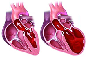
The heart is the organ responsible for pumping blood to and from all tissues of the body. The heart is divided into right and left sides. The right side of the heart pumps oxygen-deficient blood returning from the body through the lungs where it is re-oxygenated, and gets rid of the carbon dioxide waste that the body produces. After the blood passes through the lungs, it enters the left side of the heart where it is pumped out to the body though the aorta and other arteries. The picture below illustrates the pathway that blood takes as it moves though the heart. The structures shown in red contain blood that is rich in oxygen, whereas the structures shown in blue contain blood that has very little oxygen and high levels of carbon dioxide.
Blue arrows represent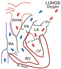 deoxygenated blood flowing through the right side of the heart to the lungs, where red arrows represent oxygenated blood leaving the lungs through the left side of the heart. RA = right atrium, RV = right ventricle, TV = tricuspid valve, PA = pulmonary artery, PV = pulmonary vein, LA = left atrium, MV = mitral valve, LV = left ventricle.
Each side of the heart has two chambers, an upper atrium and a lower ventricle. Between the atrium and ventricle on each side lies a valve – the tricuspid on the right and the mitral on the left – that regulates blood flow from the upper atrial chambers into the lower ventricular chambers. As the heart pumps (squeezes), these valves act as one-way gates allowing blood to flow from the atrium above to the ventricle below and preventing blood from flowing backwards into the atrium.
From the ventricles, blood is pumped out into the lungs through the pulmonary artery (on the right) or out to the body through the aorta (on the left) through a second series of one-way valves (the pulmonic valve on the right and the aortic valve on the left).
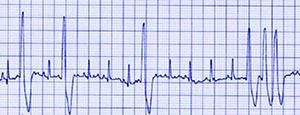
The number of heartbeats per minute (heart rate) and the type of heartbeats are controlled by the electrical system in the heart. Normal heartbeats start in the right atrium, but in sick hearts, the heart beats can start from any chamber and are called arrhythmias.
What is Chronic Degenerative Valve Disease (CVD)?
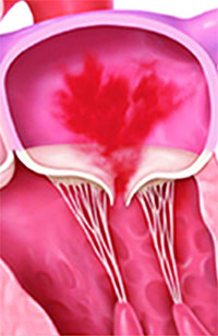
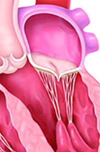
Chronic degenerative valve disease (CVD) has many other names, such as endocardiosis, valvular regurgitation, valvular insufficiency, chronic valve disease, mitral regurgitation (MR), or myxomatous degeneration of the valve. This disease is a consequence of degeneration of the valves between the atrium and ventricle on both the right (tricuspid valve) and left (mitral valve) side of the heart, however the valve on the left side (mitral valve) is typically most severely affected. The degeneration process causes the valves to become abnormally thick and develop a nodular “lumpy” appearance. This process is NOT caused by an infection. The degenerative changes in the valves and the structures that support the valves impede their ability to form a tight seal between the atrium and ventricle when the heart is squeezing or pumping This causes blood to leak backwards into the upper chambers (atria). The backwards leaking of blood through the abnormal valve is called “regurgitation” and causes an abnormal sound called a heart murmur that is typically heard with a stethoscope by your veterinarian. The consequences of CVD are discussed below.
Who develops Chronic Degenerative Valve Disease?
Chronic degenerative valve disease represents approximately 75% of all heart disease in the dog and is very, very rare in the cat. Approximately 60% of affected dogs have degeneration of the mitral valve, 30% have degeneration of both the tricuspid and mitral valves, and involvement of the tricuspid valve alone occurs in 10% of affected dogs. The risk of developing CVD increases as dogs get older and is rare before the age of 4 years. In addition, small breed dogs (dogs weighing less than 40 lb (18.2 kg) are more likely to get CVD than larger dogs. Certain breeds also have a higher risk of developing CVD.
General risk factors for the development of CVD.
- Dog
- Older (> 5 years)
- Small breed (
< 40 lb; (18.2 kg) - High risk breeds, including:
– Miniature poodles, cocker spaniels, miniature schnauzers, Dachshunds, small terrier breeds
– Cavalier King Charles spaniels (this breed is an exception in that they may develop CVD as young as 2-3 years of age)
What causes CVD?
CVD is a degenerative process associated with aging in predominantly small dogs. It is not caused by an infection. Many older dogs with dental disease also have CVD but the dental disease is not the cause of the CVD. However, good dental hygiene is an important part of ensuring that your dog lives a long and healthy life. In addition, there is likely a familial or inherited genetic component in some breeds, such as the Cavalier King Charles spaniel, however the specific genetic evidence for the majority of cases of CVD is lacking.
What happens to my dog when it gets CVD?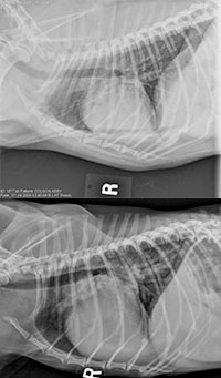
The leak (regurgitation) caused by the degeneration of the valves eventually leads to enlargement of the heart chambers (the atria and ventricles). The majority of dogs with CVD feel and act normally and the only evidence that they have CVD is the sound of the leak (heart murmur) that is heard by your veterinarian when using a stethoscope. This is typically called the preclinical stage of the disease (or Stage B1 and B2), i.e. your dog is considered ‘asymptomatic’. Most dogs with asymptomatic CVD never get sick from CVD, however about 1/3 will eventually develop ‘clinical signs’ or clues that you will notice (see list below). In general, dogs with very enlarged (the biggest) hearts (Stage B2) are more likely to develop ‘clinical signs’. The regurgitation and intensity of the heart murmur typically gets bigger and louder, slowly, over years, but sometimes these changes can happen quickly. The bigger the leak, the bigger the heart gets and eventually when the heart is big enough, your dog can start to have problems (clinical signs) that you will notice, such as fast breathing, increased effort when breathing, coughing and other clinical signs (see the list below). The clinical signs occur when the pressures in the enlarged heart chambers cause fluid to leak out of the blood vessels into the lungs (called,pulmonary edema; ‘water on the lungs’) and sometimes the belly (ascites). The development of clinical signs is referred to as the ‘symptomtic’ stage of CVD (Stage C). The build up of fluid in the symptomatic stage is called congestive heart failure. In addition, sometimes in the symptomatic stage of CVD the heart cannot send enough blood forward to the body and this can cause dogs to become weak or tired or even faint.
Clinical signs (clues) that your dog may have ‘symptomatic’ CVD and needs to be examined by your veterinarian.
Note: These clues represent clinical signs that may occur in dogs with CVD once they become ‘symptomatic’, but they can also occur with other diseases. However, if you know your dog has CVD you should always watch for their development and if they do, you should contact your veterinarian. It is important to note that not every dog will develop all of the following clinical signs and many dogs will have more than one.
Clinical signs that can be associated with CVD.
- Fast (rapid and shallow) breathing when resting or sleeping (> 30-35 breaths per minute)
- Note: For details on how and why to evaluate this in your dog, refer to the section on evaluating home breathing rates below.
- Increased effort associated with breathing
- Restless or agitation while sleeping, i.e. moving around a lot and changing positions due to the inability to find a comfortable position
- Change in the position that your dog sleeps in, i.e. if your pet no longer sleeps on its back or on its side, but more in a sitting or ‘sphinx position’.
- Coughing or gagging
- Weakness
- Reduced ability to exercise
- Collapse or fainting
- Decreased appetite
- Weight loss
- Distended belly
- Depressed attitude or quiet and not interactive
How can my veterinarian determine if my dog has CVD?
The best screening test to detect CVD in your dog is listening (auscultation) with a stethoscope by your veterinarian once per year. Other tests will be recommended if your veterinarian hears a heart murmur. If you wish to have your dog ‘cleared’ for breeding purposes your veterinarian may recommend that you have your dog auscultated by a veterinary cardiologist.
What tests might be recommended by my veterinarian once my dog has CVD?
Asymptomatic dogs with CVD:
If your dog is asymptomatic (no clinical signs of CVD are present, as outlined above) and your veterinarian detects a heart murmur , further tests will be recommended to help determine how big the heart is and if your dog should start taking any heart medications. Some tests are done to determine if there are any other problems that could be detrimental for dogs with have CVD, for example high blood pressure or kidney disease.
Symptomatic dogs with CVD:
Dogs with a heart murmur and one or more of the clinical signs listed above could have heart failure, which requires medications. Your veterinarian will recommend tests to help determine the cause of the clinical signs and help them select the appropriate medications.
Possible tests for dogs with asymptomatic and symptomatic CVD:
- Chest x-rays to evaluate the heart size, lungs and blood vessels
- Blood work to evaluate kidney function
- Urine test to evaluate kidney function
- Blood pressure
- Ultrasound (echocardiogram of the heart) to confirm the diagnosis and evaluate the size and function of the heart.
- A blood test called, NT-proBNP, to evaluate the pressure in the heart
- ECG to evaluate the heart rhythm
- Referral to a veterinary cardiologist may be suggested in some cases
How is CVD treated in dogs?
Asyptomatic CVD:
Medication is not required if the recommended tests determine that the heart is not enlarged and the blood pressure is normal. However, if the tests detect heart enlargement and/or high blood pressure, medication(s) may be prescribed.
Can I slow down or reverse the progression of CVD in my dog?
Several studies have looked at preventing CVD from progressing to the development of congestive heart failure. Unfortunately, no drugs that have been tested thus far have been proven to be effective in either preventing or slowing down the progression of CVD. As new drugs are developed, they will also undergo testing, so there may be drugs in the future that could help with CVD progression.
Symptomatic CVD (heart failure):
Medications will be prescribed if your dog has clinical signs and the results of the recommended tests have led to a diagnosis of heart failure. These medications will be continued forever, although the doses and frequency of administration may change over time. Sometimes additional medications are required, while others are sometimes stopped. In addition, your dog may need to stay in the hospital for a few days while adjusting to the medications.
Common medications used to treat heart failure due to CVD:
- Furosemide, also known as Lasix®
- Pimobendan (Vetmedin®)
- Angiotensin converting enzyme inhibitor (ACEI), such as benazepril or enalapril
- Spironolactone
- Additional medications are frequently indicated in individual dogs
Note: Some medications and herbal supplements can have adverse interactions with heart medications, therefore it is important that you not use any medications (new or previously prescribed) without talking to the veterinarian that prescribes your dog’s heart medications.
Common side-effects of medication used to treat heart failure due to CVD:
- Drinking large amounts of water more frequently
Note: It is critical that dogs on medications like furosemide have free choice access to water at all times.
- Urinating larger amounts more frequently
Note: Accidents in the house can be limited by using ‘doggy doors’ and being sure to not give the furosemide pills within 2 hours of bedtime or leaving the house for an extended period of time. Dogs always need to urinate within 1-2 hours after receiving furosemide.
- Reduced appetite or not eating normal amounts of food
Note: If this problem starts and persists, you need to talk to yourveterinarian; it is important that your dog not loose weight.
What kind of follow-up will my dog need now that it has CVD?
Recommended follow-up in dogs with asymptomatic CVD (Stage B1 and B2):
Your veterinarian will recommend recheck appointments every 3-12 months depending on how advanced the disease is. In addition, if and when a medication is started you may need to return in 10-14 days to have a blood test to check the kidney and liver values, and take your dog’s blood pressure. However, if you notice any of the clinical signs listed above at any time, you should not wait for the next recheck appointment, but call and make an urgent/ emergency appointment. If the clinical signs are severe and or develop suddenly (your dog cannot breath, rest comfortably or sleep) you may need to take him to the nearest emergency facility as soon as possible. One of the best ways to help your veterinarian determine when to start or adjust your dogs heart medication(s) is to observe and record the home resting/sleeping breathing rate of your dog (see below).
Recommended follow-up in dogs with symptomatic CVD (Stage C):
Your veterinarian will recommend a recheck appointment every 2-4 months. In addition, if and when a new medication is started you may need to return in 10-14 days to have a blood test to check the kidney and liver values, and take your dog’s blood pressure. However, if you notice any of the clinical signs listed above at any time, you should not wait for the next recheck appointment, but call and make an urgent/ emergency appointment. If the clinical signs are severe, and or develop suddenly (your dog cannot breath, rest comfortably or sleep) you may need to take him to the nearest emergency facility as soon as possible. One of the best ways to help your veterinarian determine when to start or adjust your dogs heart medication(s) is to observe and record the home resting/sleeping breathing rate of your dog (see below).
Note: Please see the “Measuring Your Pet’s Breathing Rate” brochure in this series for more information on how to monitor your pet’s breathing rate at home.
Can special diets help dogs with CVD live longer?
Some animal diet manufacturers have developed heart-specific diets. Some of these diets are severely restricted in salt and some are moderately restricted in salt. The diets that are severely restricted in salt should never be used in asymptomatic dogs with CVD. They may be used in dogs with heart failure as long as the dog will eat them. The heart-specific diets that are moderately restricted in salt (or any diet that is moderately restricted in salt, such as most senior diets) can be used in asymptomatic dogs with CVD. However, while these diets are unlikely to be harmful if used as outlined above, they have not been shown to have any benefit. Therefore, it is unlikely that your veterinarian will recommend that you change your dog’s diet if it is otherwise healthy. It is important that dogs and cats with advanced heart disease continue to eat.
Note: It is often beneficial to limits treats that are high in sodium in dogs that have heart disease especially those in heart failure.
Are there any dietary supplements that may help dogs with CVD live longer?
Supplements are unlikely to be harmful if used as outlined by a veterinarian who is familiar with all the medication your dog is receiving and what type and severity of heart disease your dog has However, there is no proof that your dog needs to take any supplement(s) if they are on a high quality commercial dog food diet. The most common supplement recommended in dogs with CVD is omega 3 fatty acids. You should discuss any supplements your dog receives, or any new ones you wish to start, with your veterinarian.
What about exercise in my dog once it has CVD?
In general, dogs with both asymptomatic and symptomatic CVD should be allowed to exercise at their normal level if they want to. However, the duration of sustained strenuous activities such as ball retrieval, swimming, Frisbee etc. should be limited especially in really hot or cold weather. Some exercise is good for you and your dog and part of what helps your dog enjoy their life.
Is there a surgical treatment option for dogs with CVD?
In human medicine, it is common for patients with significant CVD to have the leaky valve(s) repaired or replaced by a surgeon. However this requires openheart surgery and cardiopulmonary bypass and dogs, especially small dogs, are very hard to perform cardiopulmonary bypass on. Thus, open-heart surgery and valve replacement or repair is not readily available in the dog. A few veterinary surgeons have performed these surgeries on a very limited number of dogs. There is a very high risk of death and the surgery is very expensive (at least $10,000),therefore this option is not recommended at this time. If you wish to investigate the possibility of surgical repair or replacement, you should discuss this with your veterinarian. In the future, catheter-based (minimally invasive) techniques may become available and allow repair or replacement to be done safely without the need for cardiopulmonary bypass. Heart transplantation is not an option in dogs.
How long will my dog live now that they have CVD?
Asymptomatic CVD (Stage B1 and B2):
On average, the majority of dogs with asymptomatic CVD will live for many years (1-5 years or longer) without ever developing any clinical signs of heart failure. It can be difficult to determine the rate of progression of CVD in individual dogs, and it is for this reason that your veterinarian recommends tests and recheck appointments. The amount of change in heart size and other factors between recheck appointments can help your veterinarian determine the how your dog is doing.
Symptomatic CVD (Stage C or heart failure):
Once your dog develops clinical signs of CVD, medications can typically eliminate or reduce the severity of the clinical signs but the disease will still progress and eventually the medications will not work as well. Sometimes, new medications can be added or the doses of your dogs’ current medications adjusted but eventually this will be insufficient in maintaining your dog’s quality of life. Your veterinarian will help you recognize when and if this is the case for your dog. However, in general, with the appropriate medications and recheck appointments, many dogs with heart failure due to CVD live for 1-2 years or even longer.
Dialated Cardiomyopathy (DCM)
Download the “Dialated Cardiomyopathy (DCM)” information pamphlet (PDF).
How does the normal heart work?
The heart is the organ responsible for pumping blood to and from all tissuesof the body. The heart is divided into right and left sides. The job of the right side is to pump oxygen-deficient blood returning from the body through the lungs to be reoxygenated, and to get rid of the carbon dioxide waste that the body produces. After the blood passes through the lungs and picks up oxygen and gets rid of the carbon dioxide waste it enters the left side of the heart where it is pumped out to the body though the aorta and other arteries. The picture below illustrates the pathway that blood takes as it moves though the heart. The structures shown red contain blood that has lots of oxygen whereas the structures shown in blue contain blood that has very little oxygen and high levels of carbon dioxide.
Blue arrows represent deoxygenated blood flowing through the right side of the heart to the lungs, where red arrows represent oxygenated blood leaving the lungs through the left side of the heart.
RA = right atrium, RV = right ventricle, TV = tricuspid valve, PA = pulmonary artery, PV = pulmonary vein, LA = left atrium, MV = mitral valve, LV = left ventricle.
Each side of the heart has two chambers, an upper atrium and a lower ventricle. Between the atrium and ventricle on each side lies a valve – the tricuspid on the right and the mitral on the left – that regulates blood flow into the lower ventricular chambers. As the heart pumps (squeezes), these valves act as one-way gates allowing blood to flow from the atrium above to the ventricle below and preventing blood from flowing backwards into the atrium when the ventricle squeezes. From the ventricles, blood is then forced to flow out into the lungs through the pulmonary artery (on the right) or out to the body through the aorta (on the left) through a second series of one-way valves (the pulmonic valve on the right and the aortic valve on the left). The number of heartbeats per minute (heart rate) and the type of heartbeats is controlled by the electrical system in the heart. Normal heartbeats start in the right atrium, but in sick hearts, heartbeats can start from any chamber.
What is dilated cardiomyopathy (DCM)?
DCM is a disease that causes the heart muscle cells (myocardium) to become weak and frequently causes abnormal heart beats (an arrhythmia)  to occur. Both of these problems can cause the heart to lose its ability to contract or pump blood effectively out to the body. DCM can affect both sides of the heart simultaneously or separately but typically it affects the left sided chambers, especially the left ventricle. DCM eventually leads to two main types of problems that can occur alone or together. First, because the heart muscle is weak and does not pump blood effectively, blood backs up in the heart chambers and causes them to enlarge or dilate. In some dogs that have DCM when the heart is significantly dilated the mitral (+/- tricuspid) valves can leak and cause a heart murmur that your veterinarian can hear with a stethoscope. Second, abnormal heartbeats can occur intermittently though-out the day and night and can reduce the amount of blood pumped to the body and this can lead to weakness or fainting or even sudden death. In cases where the heart chamber size and pumping function are still normal but abnormal heartbeats are present DCM is often referred to as the arrhythmogenic form of DCM. In some dogs that have DCM your veterinarian may hear abnormal heartbeats or abnormal heart sounds with a stethoscope. Many dogs with DCM do not have heart murmurs, abnormal heartbeats or abnormal heart sounds when your veterinarian listens with a stethoscope.
What types of dogs does DCM affect?
DCM is a relatively common heart disease in the dog accounting for approximately 10% of all heart disease. Large and giant breeds are most commonly affected. DCM is very rare in smaller breed dogs (
< 30lb or 13.6kg and cats. The risk of getting DCM increases as dogs get older and rarely occurs in dog that are < 4 years old. Certain breeds have a higher risk of developing DCM, especially Doberman Pinschers.
General risk factors for DCM:
- Dog
- Older (>4 years)
- Large or giant breed (>30 lb or 13.6 kg)
- High-risk breeds, including:
- Doberman Pinscher (Approximately 60% chance of developing DCM at some point during their life)
- Cocker Spaniels
- Boxers (May get the arrhythmogenic form of DCM-chamber size and pump function is normal but abnormal heart beats are present-as young as 1 year of age
- Great Danes
What are the causes of DCM?
There are many theories as to the cause of DCM, however the true cause has not been identified and is often referred to as idiopathic for that reason. A familial or inherited genetic component is believed to exist in most cases. However, specific genetic evidence for the majority of DCM cases is lacking.
DCM may be related to a nutritional deficiency in taurine in some dogs and cats but this is a very rare cause of DCM. Taurine is an amino acid required for the development and function of the heart muscle cells. Therefore, some pets (especially Cocker spaniels) may develop DCM if they eat taurine deficient diets or if they cannot absorb taurine normally from the food they eat.
Note: The majority of cases of DCM that are supplemented with taurine will NOT improve. However, if your pet is diagnosed with DCM, ask your veterinarian if they feel that your pet would benefit from taurine supplementation.
L-carnitine is another amino acid that is required for the heart muscle cells to produce energy and pump normally. A nutritional deficiency in L-carnitine can also lead to the development of DCM in some dogs (especially some Boxers and Cocker spaniels). However, the role of L-carnitine deficiency in most dogs with DCM is unknown.
Note: The majority of cases of DCM that are supplemented with L-carinitne will NOT improve. However, if your pet is diagnosed with DCM, ask your veterinarian if they feel that your pet would benefit from L-carnitine supplementation.
What happens to my dog when it gets DCM?
There are two main forms of cardiomyopathy in the dog. The first causes the heart muscles to get weak. This causes blood to back-up into the heart chambers and then the heart chambers enlarge or dilate. The second form causes abnormal heartbeats to occur. Dogs with DCM can have one form at time or suffer from both forms at the same time. Early in the course of the disease dogs with DCM feel and act normal and the only evidence that they have DCM may be the fact that your veterinarian heard a heart murmur, abnormal heartbeat(s) or an abnormal heart sound. In some cases DCM may be diagnosed because your dog underwent a screening test, such as an echocardiogram or Holter recorder, because they are considered to be a high-risk breed such as a Doberman or a Boxer. At this stage DCM is typically called asymptomatic, preclinical or occult (Stage B). The majority of dogs with asymptomatic DCM will develop ‘clinical signs’ 6-24 months after they are diagnosed
  with asymptomatic DCM. In general, the dogs with the biggest (most dilated) hearts and the dogs with the most abnormal heartbeats are the most likely to develop ‘clinical signs’. The clinical signs or clues that you might notice if your dog has DCM include: fast breathing, increased effort when breathing, coughing or fainting (see the list below). In some cases the first sign that a dog has DCM could be that they die suddenly. The problems or ‘clinical signs’ occur when the pressures in the dilated heart chambers cause fluid to leak out of the blood vessels into the lungs (called pulmonary edema) and sometimes the belly (ascites) or when there are a lot of abnormal heartbeats. Once a dog develops clinical signs it is considered to have symptomatic DCM. The build up of fluid in the symptomatic stage is called congestive heart failure (Stage C).
Clinical signs that may be associated with DCM in dogs, include:
Note: These clues represent ‘clinical signs’ that may occur in dogs with DCM but can also occur with other diseases. However, if you know your dog has DCM you should always watch for them to develop and if they do you should contact your veterinarian. Not every dog will develop all of the following ‘clinical signs’ and many dogs will have more than one.
- Fast breathing when resting or sleeping (> 30-35 breaths per minute)
- Note: for details on how and why to evaluate this in your dog see the section on evaluating home breathing rates below
- Increased effort associated with breathing
- Restless sleeping, moving around a lot and changing positions
- Coughing or gagging
- Weakness
- Reduced ability to exercise
- Collapse or fainting
- Decreased appetite
- Weight loss
- Distended belly
- Depressed attitude or quiet and not interactive
- Sudden death
How can my veterinarian determine if my dog has DCM?
Because the majority of dogs with asymptomatic DCM are normal and often have no abnormalities that can be detected by your veterinarian with a stethoscope they frequently do not get diagnosed with DCM until they are symptomatic. In some high risk breeds such as the Boxer and Doberman pinscher your veterinarian may recommend annual screening tests to determine if DCM in present even if you dog is apparently healthy.
Screening tests for DCM that my veterinarian may recommend:
Note: Screening tests are typically only recommended in high-risk breeds and some of these tests may require referral to a veterinary cardiologist or other veterinary specialist. Other tests maybe recommended if DCM is diagnosed (see below).
- Echocardiogram-ultrasound of the heart
- Holter recorder-24 hr ECG that your dog wears home to detect abnormal heart beats
- ECG-3 minute evaluation that is done in the hospital to detect abnormal heart beats
- NT-proBNP-might be used in Dobermans to help determine if your Doberman should have an echocardiogram
- Genetic tests-might be used in combination with other screening tests
Note: These genetic tests are only available for certain high-risk breeds such as the Doberman and Boxer. It is important to know that a positive genetic test does not mean that your dog has DCM or will get DCM and a negative test does not mean they cannot get DCM. These tests are typically most useful for breeders to help plan breeding programs.
What tests might be recommended by my veterinarian once my dog has DCM?
Asymptomatic dogs with DCM:
If your dog is asymptomatic (no clinical signs of DCM are present as outlined above) and your veterinarian detects a murmur, abnormal heart beats or an abnormal heart sound with a stethoscope they will recommend test(s) to help them determine it DCM is present and to determine if your dog should start taking any heart medications. Some tests are done to determine if there are any other problems that would be bad for dogs to have if they have DCM, for example high blood pressure or kidney disease.
Symptomatic dogs with DCM (heart failure and or abnormal heartbeats):
Dogs with one or more of the clinical signs listed above could have heart failure and require medications. Your veterinarian will recommend tests to help determine the cause of the clinical signs and help them select the appropriate medications.
- Chest radiographs identify any pulmonary edema (fluid in the lungs backed up from the heart) and evaluate the heart size
- Echocardiogram-ultrasound of the heart to confirm the diagnosis and evaluate the size and function of the heart
- ECG identify abnormal heart beats
- Holter recorder-24 hour ECG that the dog wears home to help evaluate abnormal heartbeats
- Blood tests evaluate kidney and liver function
- Urine test evaluates kidney function
- Thyroid blood test
- Blood pressure
- NTproBNP to evaluate the pressure in the heart
- Genetic testing is available for the in the Boxer and Doberman but does not replace other important tests and cannot diagnose DCM
- Referral to a veterinary cardiologist may be suggested in some cases
How is DCM treated in dogs?
The treatment of DCM depends on the stage of the disease and the individual dogs problems.
Asymptomatic dogs with DCM:
There are 3 medications that are commonly used to treat asymptomatic DCM; pimobendan (Vetmedin), an angiotensin converting enzyme inhibitor (enalpril or benzepril), and an antiarrhythmic (sotalol). These medications can be used alone or together and in some cases other medications may also be prescribed. The goal of these medications is to slow down the progression of the disease and help your dog stay asymptomatic and live longer.
Symptomatic dogs with DCM?
Medications will be prescribed if your dog has clinical signs and the results of the recommended tests have led to a diagnosis of heart failure with or without a large number or abnormal heart beats. These medications will be continued forever, although the doses and frequency of administration may change over time. Sometimes additional medications are required, while others are sometimes stopped. In addition, your dog may need to stay in the hospital for a few days while adjusting to the medications.
Common medications used to treat heart failure and abnormal heart beats due to DCM:
- Furosemide, also known as Lasix®
- Pimobendan (Vetmedin®)
- Angiotensin converting enzyme inhibitor (ACEI), such as benazepril or enalapril
- Spironolactone
- Sotalol, for abnormal heartbeats
- Mexilitine, for abnormal heartbeats
- Diltiazem, for abnormal heartbeats
- Additional medications are frequently indicated in individual dogs
Note: Some medications and herbal supplements can have adverse interactions with heart medications; therefore it is important that you not use any medications (new or previously prescribed) without talking to the veterinarian that prescribes your dog’s heart medications.
Common side-effects of medication used to treat heart failure and or abnormal heart beats due to DCM, include:
- Drinking large amounts of water more frequently
- Note: It is critical that dogs on medications like furosemide have free choice access to water at all times.
- Urinating larger amounts more frequently
- Note: Using ‘doggy doors’ and being sure to not give the furosemide pills within 2 hours of bedtime or leaving the house for an extended period of time can limit accidents in the house. Dogs always need to urinate within 1-2 hours after receiving furosemide.
- Reduced appetite or not eating normal amounts of food
- Note: If this problem starts and persists, you need to talk to your veterinarian; it is important that your dog does not loose weight.
What kind of follow-up will my dog need now that it has DCM?
Recommended follow-up in dogs with asymptomatic DCM (Stage B):
 Your veterinarian will recommend recheck appointments every 3-6 months depending on how advanced the disease is. In addition, if and when a medication is started you may need to return in 10-14 days to have a blood test to check the kidney and liver values, take your dog’s blood pressure or re-evaluate the abnormal heart beats. However, if you notice any of the clinical signs listed above at any time, you should not wait for the next recheck appointment, but rather call and make an urgent/emergency appointment. If the clinical signs are severe and or develop suddenly (your dog cannot breath, rest or sleep comfortably Normal Heart Dilated Cardiomyopathy Dilated ventricle or collapses or faints) you may need to take them to the nearest emergency facility as soon as possible. One of the best ways to help your veterinarian determine when to start or adjust your dogs heart medication(s) is to observe and record the home resting/sleeping breathing rate of your dog (see below).
Recommended follow-up in dogs with symptomatic DCM (heart failure and or abnormal heartbeats):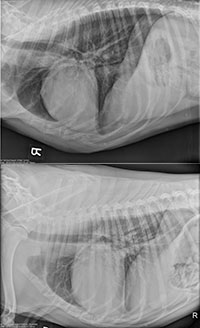
Your veterinarian will recommend a recheck appointment every 2-4 months. In addition, if and when a new medication is started you may need to return in 10-14 day to have a blood test to check the kidney and liver values, and take your dog’s blood pressure.
However, if you notice any of the clinical signs listed above at any time, you should not wait for the next recheck appointment, but rather call and make an urgent/emergency appointment. If the clinical signs are severe, and or develop suddenly (your do cannot breath, rest or sleep comfortably or collapses or faints) you may need to take them to the nearest emergency facility as soon as possible. One of the best ways to help your veterinarian determine when to start or adjust your dogs heart medication(s) is to observe and record the home resting/sleeping breathing rate of your dog (see below).
Why should I evaluate my dog’s resting/sleeping breathing rate at home?
An increase in resting or sleeping breathing rate is an important early clinical sign of heart failure that you can evaluate at home.
This is an early clue that heart failure is developing, and your observations can help limit how sick your dog becomes, reduce the chances that your dog will ever have to stay overnight in the hospital, and therefore also help reduce the costs associated with heart failure treatment.
Note: Please see the “Measuring Your Pet’s Breathing Rate” brochure in this series for more information on how to monitor your pet’s breathing rate at home.
Can special diets help dogs with DCM live longer?
Some animal diet manufacturers have developed heart-specific diets. Some of these diets are severely restricted in salt and some are  moderately restricted in salt. The diets that are severely restricted in salt should never be used in asymptomatic dogs with DCM. They may be used in dogs with heart failure as long as the dog will eat them. The heart-specific diets that are moderately restricted in salt (or any diet that is moderately restricted in salt, such as most senior diets) can be used in asymptomatic dogs with DCM. However, while these diets are unlikely to be harmful if used as outlined above, they have not been shown to have any benefit. Therefore, it is unlikely that your veterinarian will recommend that you change your dog’s diet if it is otherwise healthy.
Note: It is often beneficial to limit treats that are high in sodium in dogs that have heart disease, especially those in heart failure.
Are there any dietary supplements that may help dogs with DCM live longer?
Supplements are unlikely to be harmful if used as outlined by a veterinarian who is familiar with all the medication(s) your dog is receiving and what type and severity of heart disease your dog is suffering from. However, in most cases there is no proof that your dog needs to take any supplement(s) if they are on a high quality commercial dog food diet. The most common supplement recommended in dogs with DCM is omega 3 fatty acids and in some cases taurine and or L-carnitine. You should discuss any supplements your dog receives, or any new ones you wish to start, with your veterinarian.
What about exercise for my dog once it has DCM?
In general, dogs with both asymptomatic and symptomatic DCM should be allowed to exercise at their normal level if they want to. However, the duration of sustained strenuous activities such as ball retrieval, swimming, Frisbee etc. should be limited especially in really hot or cold weather. Some exercise is good for you and your dog and part of what helps your dog enjoy their life.
Are there any research studies being conducted to learn how to treat DCM more effectively?
Some researchers (Dr. Amara Estrada at the University of Florida Veterinary School) are evaluating stem cell treatment in Dobermans. In future gene therapy may also be available for some dogs. Heart transplantation is not an option in dogs.
How long will my dog live now that it has DCM?
Asymptomatic dogs with DCM:
On average, the majority of dogs with asymptomatic DCM can live for 1-3 years before they develop any clinical signs of heart failure. However, a few will die suddenly. This can happen at any time but is most common in the dogs that have a lot of abnormal heartbeats and is not painful for your dog. It is however difficult to determine the evolution of an individual, and it is for this reason that your veterinarian recommends tests and recheck appointments.
Symptomatic dogs with DCM:
Once your dog develops clinical signs of DCM, medications can typically eliminate or reduce the severity of the clinical signs but the disease will still progress and eventually the medications will not work as well. In some cases, your dog will seem to be doing very well but will die suddenly. This is most common in dogs with most common in the dogs that have a lot of abnormal heartbeats and is not painful for your dog. Sometimes, new medications can be added or the doses of your dogs’ current medications adjusted but eventually this will be insufficient in maintaining your dog’s quality of life. Your veterinarian will help you recognize when and if this is the case for your dog. However, in general, with the appropriate medications and recheck appointments, many dogs with heart failure due to DCM live for more than six-12 months.
Heartworm Disease: Prevention and Treatment
Download the “Heartworm Disease: Prevention and Treatment” information pamphlet (PDF).
WATCH: The life cycle of the heartworm parasite.
What is heartworm disease?
Heartworm disease (HW) is a potentially serious problem in dogs in many geographic areas including almost all of the United States. Years ago HW was only common in the southern coastal areas of the U.S., but due to the movement of dogs with the human population, the disease has spread over most of the country. However, with proper preventive therapy, heartworm disease will not be a risk to your dog.
HW is caused by Dirofilaria immitis, a parasitic worm that lives as an adult predominantly in large blood vessels in the lungs and in some cases in the right side of the dog’s heart. Heartworms do most of the damage to the heart and lungs in the adult stage, at which time the worms can measure over one foot long. Mosquitoes transmit heartworms from dog to dog. The adult heartworms can live in a dog for up to 7 years. The adults produce microfilariae, or heartworm larvae, which can be found within the bloodstream of affected dogs if adult female worms are present in their lungs.
What are the clinical signs of HW?
Signs of HW may occur within six months of infection (after a dog is bitten by an infected mosquito) or may not appear at all. Whether or not a dog develops clinical signs depends on the severity of the infection (number of adult worms that are present) and the dog’s own response to the worms. In most cases, signs will begin within 1-2 years after infection. Typical signs include coughing, labored breathing, weakness, and tiring with exercise. Since the signs vary, the disease may be advanced before a dog begins to show any signs of infection. In some dogs, signs may be mistaken for another problem. In advanced disease, the heart and lungs can be severely damaged or even plugged up with worms. Eventually, heart failure can occur and dogs can die from damage caused by heartworms before, during, or after appropriate treatment is started.
Clinical signs that may be associated with HW infection in dogs, include:
- Coughing
- Labored breathing
- Fast breathing
- Weakness
- Tiring with exercise
- Collapse or fainting
- Weight loss
- Dark colored urine (color of red wine)
- Distended abdomen
Note: These signs may occur in dogs with HW but can also occur with other diseases. Not every dog will develop all of the following ‘clinical signs’ and many dogs will have more than one.
How is HW diagnosed?
Typically, a blood sample will be taken from your dog to test for heartworm infection. This test detects markers (antigens) from adult female worms and is very accurate. Chest radiographs (x-rays), blood tests to evaluate the kidneys and liver, and/or an echocardiogram (ultrasound of the heart) may also be recommended in some cases.
What are the treatment options for HW?
The most common treatment for HW requires that your veterinarian give an injectable medication to kill the adult heartworms. Currently, there is only one medication available for the treatment of adult heartworms. The medication is called melarsomine (Immiticide®) and is an arsenical type compound that kills the adult heartworms. It is the treatment recommended by the American Heartworm Society. There are two basic treatment options with Immiticide®-the two-injection method and the three-injection method. The American Heartworm Society recommends the three-injection method for all dogs. Following treatment with each injection, your dog must be STRICTLY rested for four to six weeks, during which time the dead adult heartworms will be slowly destroyed and removed by the body. Exercise increases blood flow within the heart and lungs and commonly leads to significant complications after treatment, such as pieces of dead worms moving deeper into the lungs causing coughing, severe difficulty breathing, distress, and even sudden death. Preventive medication for HW is also started during treatment to prevent new infections.
Surgical removal of heartworms is recommended when dogs have a large number of heartworms in their heart chambers. This condition is often called ‘caval syndrome’ and can develop suddenly in some dogs. Heartworm removal in these cases involves making a small incision in the skin on the neck and also in the jugular vein that leads to the heart. Heartworms are manually removed with special steerable, bendable forceps or a basket. Within a few weeks following recovery from surgery, standard injectable treatment is recommended to eliminate any remaining worms in the lungs. Worms in the lungs cannot be removed manually.
How is HW prevented?
To prevent heartworm infection in the dog population and to provide your pet with the best possible protection against heartworms, the following guidelines are recommended:
- All puppies should be started on preventive medication no later than eight weeks of age.
- Your dog should be evaluated routinely (typically once a year) for heartworms, particularly with an antigen blood test. This is especially important if there are occasional times your dog has not received its HW prevention medication.
- All dogs that live in high risk areas or travel to high risk areas should be on preventive edication year round. In some areas (cooler climates), your veterinarian may recommend that you only need to use the preventative medication during the high-risk season when mosquitos are around. However, the Heartworm Society of North America recommends year-round preventative medication for all dogs.
- Dogs that show new signs of heartworm disease as outlined above should be have a HW antigen blood test performed.
- If your dog is diagnosed with HW, a veterinarian should treat to limit, and ideally, prevent permanent damage to the heart and lungs, as well as to keep your dog alive.
- Prevention is the best approach to HW. If preventive medication and monitoring programs are followed, unnecessary treatment, permanent heart and lung damage, and heartworm-related death can be avoided.
Measuring Your Pet’s Breating Rate
Download the “Measuring Your Pet’s Breathing Rate” information pamphlet (PDF).
 Why should I evaluate my pet’s breathing rate at home?
Increases in your pet’s breathing rate while resting quietly or sleeping is a very important early clue (‘clinical sign’) that your pet may be developing heart failure and needs to see your veterinarian. Since this is an early clue that heart failure is developing, by noticing you can help limit how sick your pet will get, reduce the chances that your pet will ever have to stay overnight in the hospital, and therefore also help reduce the costs associated with heart failure treatment.
What is a normal resting/sleeping breathing rate for dogs and cats?
In general, all dogs and cats, with or without heart disease, have a breathing rate of between 15-30 breaths every minute. Lower rates are even possible and are no cause for concern as long as your pet is otherwise healthy. Breathing rates are much higher than this when dogs and cats are hot, stressed or active but that is OK.
Resting/sleeping breathing rates that are consistently greater than approximately 30 per minute are increased and abnormal. In some cases rates lower than 30 per minute may be considered increased and abnormal by your veterinarian. You should ask your veterinarian what rate is considered increased and abnormal for your dog or cat.
Clinical signs that may be associated with heart disease or heart failure in dogs and cats include:
- Fast breathing when resting or sleeping (> 30 breaths per minute)
- Increased effort associated with breathing
- Restless sleeping, moving around a lot and changing positions
- Coughing or gagging
- Weakness
- Reduced ability to exercise
- Collapse or fainting
- Decreased appetite
- Weight loss
- Distended belly
- Depressed attitude or quiet and not interactive
Cat only:
- Hind leg lameness or weakness
- Hind end paralysis
- Pain
How do I count the resting/sleeping breathing rate in my pet?
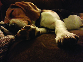
Wait until your pet is sleeping quietly (preferred) or resting calmly and quietly. It is important that cats not be purring when you count their breathing rate. The sleeping breathing rate is typically a little lower than the resting breathing rate. Then watch the chest. It moves in and out as dogs and cats breathe. One breath is counted when the chest has moved in and out once. Use your watch or phone to time 30 sec then count how many breaths occur in 30 sec. Next multiply the number of breaths that you counted in 30 sec by 2 to get the number of breaths in 60 sec or 1 minute.
Alternatively, you can count the total number of breaths that occur in 60 sec (1 min) and then there is no need to multiply. Next you need to keep a record of the breathing rates you count by writing them down somewhere such as on your calendar.
VIDEO: How to measure a resting respiratory rate in a dog
What should I do if the resting/sleeping breathing rate is increased in my pet?
The first thing to do is to count it a few times over the next couple of hours to be sure it is a consistent finding. If the breathing rate is consistently increased then you need to contact your veterinarian. Typically your veterinarian will recommend a recheck appointment in the next day or two so medications can be adjusted.
Note: If the resting/sleeping breathing rate is increased and other ‘clinical signs’ as outlined above are also observed then the situation may represent an emergency. In this case, especially if it is after hours you may need to go to a veterinary emergency center.
How often should I count the resting/sleeping breathing rate in my pet?
Typically your veterinarian will have you count the breathing rate once per day for a week when you are learning so that you get comfortable doing it. This way you and your veterinarian can also learn your pets’ actual resting/sleeping breathing rate.
If your pet has asymptomatic heart disease:
Home breathing rates need not be evaluated in all pets with asymptomatic heart disease. Your veterinarian will tell you if and when it is time to start doing this in your pet. In general it is most important to start in pets with advanced asymptomatic heart disease that have a high risk of developing heart failure within the next year. In this case breathing rates are typically recorded once or twice per week although sometimes your veterinarian may ask you to do it once per day.
If your pet has heart failure:
Home breathing rate should be evaluated once per day in all pets that have heart failure and are now taking medications such as furosemide.
Where can I find free smartphone apps for home breathing rate?
There are free smartphone apps for the iPhone, Droid, BlackBerry that can help you keep track of your dog’s home breathing rate. Search the app store for “your dog’s heart resting breathing rate.”
Pacemakers
Download the “Pacemakers” information pamphlet (PDF).

Why does my dog need a pacemaker?
Most dogs that require a pacemaker have slow heart rates and clinical signs of exercise intolerance, collapse or fainting and heart failure.
What happens if my dog has a slow heart rate but we decide not to put in a pacemaker?
Clinical signs of exercise intolerance, collapse and heart failure may persist or worsen without pacemaker implantation. Occasionally, the heart will stop beating resulting in sudden death.

How is a pacemaker placed?
The pacemaker is typically placed through the jugular vein in the right side of the neck. Occasionally, the pacemaker may be placed in the abdomen if your dog has skin disease or any other disease that prevents placement in the neck. The decision on where to place the pacemaker will be decided by the doctor caring for your dog. Some dogs need one pacing lead, while others will require multiple pacing leads depending on the heart problem.
Is my dog too old for a pacemaker?
Many dogs that require a pacemaker are older. Often times, owners may attribute slowing down to aging when, in fact, it is due to a slow heart rate. Important diseases of other organ systems, however, may preclude placement of a pacemaker.

How often are recheck examinations recommended?
Rechecks to evaluate pacemaker function and battery life should be performed one month following pacemaker placement and then every six months, ideally at Texas A&M. Additional rechecks are recommended earlier if your dog exhibits weakness or collapses.

Will my dog need heart medication?
Most dogs will not require medication following pacemaker placement. However, concurrent or underlying disease may necessitate additional medical therapy.

What is needed once my dog is home from surgery?
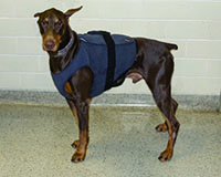
It is important that the incision be kept clean and dry. Activity should be kept to a minimum for 30 days until the incision has completely healed, and to minimize the chance of pacemaker lead dislodgement. A special diet is not required unless recommended by the doctor caring for your dog. If your dog’s pacemaker was placed in the neck, a harness should be used instead of a collar to walk your dog to prevent unnecessary pulling on the pacemaker.
What things should dogs with pacemakers avoid?
Microwaves do not present a danger to your dog. If your dog has a pacemaker and requires a Magnetic Resonance Imaging (MRI) or Computed Tomography (CT) scan, notify your veterinarian.
Patent Ductus Arteriosus (PDA)
Download the “Patent Ductus Arteriosus” information pamphlet (PDF).
")
What is a Patent Ductus Arteriosus?
Patent ductus arteriosus (PDA) is one of the most common congenital heart defects in dogs. PDA occurs more frequently in certain breeds of dogs including poodles, Shetland sheepdogs, collies, German shepherds, Maltese, Bichon Frise, Chihuahuas, Pomeranians and Newfoundlands. It is present more commonly in females than males.
The ductus arteriosus is a blood vessel that is normally present in a puppy before it is born. At birth or within a few days of birth, the ductus arteriosus should close allowing blood to begin flowing through the lungs to pick up oxygen. If the ductus arteriosus does not close, it results in an abnormal communication between the two largest vessels in the heart, the aorta and the pulmonary artery. This causes an increase in the amount of blood that flows through the left side of the heart, resulting in extra work for the left side of the heart. The increased volume of blood and extra work can cause heart failure.

How is a PDA diagnosed?
Blood flowing through the PDA causes a characteristic heart murmur typically heard at your puppy’s first visit to the veterinarian. X-rays of the chest may show heart enlargement. An echocardiogram is required to definitively diagnose PDA.
What are the clinical signs of a PDA?
Exercise intolerance, cough and breathing difficulty are the most common signs. Many dogs have no clinical signs at the time of initial diagnosis.
What are the treatment options for a PDA?
Interventional Catheterization Procedures:
With these minimally-invasive procedures, a small incision is made in the right inner-thigh and a catheter is placed into an artery in the leg. The catheter is advanced into the aorta and PDA. A dye injection is made (angiogram) allowing precise determination of the size and shape of the PDA. Then a device (coil, plug, or canine duct occluder) is placed into the PDA to stop blood flow. Occasionally, the PDA is a shape (short or tubular) that precludes the use of interventional catheterization procedures.

Surgical Ligation:
This procedure is performed by making an incision in the left side of the chest between the ribs. The heart is exposed, and the PDA is tied off to stop all abnormal blood flow. Surgery patients require pain medication after surgery, and may need an extra day of hospital care.
Both procedures typically take an average of two hours to perform. Dogs that present clinical signs of heart failure (breathing difficulty, weakness) are at a greater risk of having complications with either procedure.
What happens if the PDA is not fixed?
If not corrected, dogs with PDA have a 60% mortality in the first year. Occasionally, adult dogs are diagnosed with a PDA that is small and only caused minimal changes to the heart.
Are there any complications with either procedure?
Both procedures are typically safe (98% survival), but complications can arise. Complications include rupture and excessive bleeding of the PDA or artery in the leg, incomplete closure of the PDA, device embolization to the lungs, puncture of the heart or vessels and residual air in the thoracic cavity necessitating placement of chest tubes.
Does a dog’s size make a difference as to which procedure can be performed?
A dog’s size must be taken into consideration when determining which procedure to perform. Dogs must weigh at least 4-5 pounds in order to perform an interventional catheterization procedure. Occasionally, a small dog can wait until it is large enough to undergo an interventional catheterization procedure. This is only considered if the PDA is thought to be small, and there is no clinical evidence of important heart changes.
How do interventional catheterization procedures work?
During these procedures, one of several different devices is deployed in the PDA resulting in physical obstruction of the PDA. The device is left in place for the duration of the patient’s life, and has not been associated with any long-term complications.

What special care is needed once my dog is home from surgery?
It is important that the incision(s) be kept clean and dry. Do not allow your dog to lick or chew at the incisions. An Elizabethan collar should be placed around the neck to prevent licking or chewing, especially if the dog is going to be left alone. Do not allow your dog to play and rough house until the incision(s) are completely healed. This takes about 10-14 days. The sutures are typically buried beneath the skin, and will dissolve on their own. Occasionally, they will need to be removed by your veterinarian. A special diet is not required unless indicated by your doctor.
Will my dog’s heart return to normal?
In most cases, if clinical signs were present, they improve dramatically once the PDA is closed. The heart may return to normal size with time , but enlarged hearts may not completely return to normal. A recheck echocardiogram of the heart is recommended 1-3 months after surgery to monitor heart size and function.
Why is there a heart murmur present after surgery?
In dogs with big hearts, the mitral valve leaflets in the left side of the heart stretch apart causing a leak called mitral regurgitation. This will result in a murmur that is often heard even after the PDA is closed, and that may go away with time as the heart returns to normal size. In a small number of patients, this type of murmur may persist indefinitely.
Should a dog with a PDA be used for breeding purposes?
PDA is considered a heritable disease that can be passed to puppies.
Pulmonic Stenosis (PS)
Download the “Pulmonic Stenosis” information pamphlet (PDF).

What is pulmonic stenosis?
Pulmonic stenosis (PS) is a congenital heart defect commonly found in certain dog breeds including the English and French bulldog, boxer, miniature schnauzer, West Highland white terrier, Chihuahua and mastiff.
Normally, the pulmonic valves have three thin leaflets of tissue which close to form a tight seal. When blood is pumped out of the right side of the heart, the three leaflets move out of the way to allow the blood to pass.
The most common form of pulmonic stenosis occurs when the three leaflets are thickened and fused along their borders causing an obstruction to normal blood flow. In some dogs, the ring of tissue surrounding the pulmonic valve leaflets is too narrow. This is called annular hypoplasia.
Rarely, dogs will have a narrowing in the pulmonary artery above the pulmonic valve leaflets called supravalvular pulmonic stenosis.
How is pulmonic stenosis diagnosed?
Dogs with pulmonic stenosis have a characteristic murmur that is typically heard at your puppy’s first visit to the veterinarian. The obstruction of blood flow through the abnormal valve leaflets or narrowed tissue causes the murmur. X-rays and ultrasound (echocardiogram) show enlargement of the right side of the heart. An echocardiogram is required to diagnose the severity of pulmonic stenosis. Dogs with mild to moderate pulmonic stenosis do not typically develop clinical signs or require an intervention.

What are the clinical signs?
Commonly, puppies with pulmonic stenosis will not have any clinical signs. Some dogs have exercise intolerance. Collapse, also called syncope, may occur with excitement or exercise, and is similar to fainting. In severe cases, the right side of the heart will fail causing the abdomen to become distended with fluid.
What are the treatment options for pulmonic stenosis?
Dogs with severe pulmonic stenosis and clinical signs may benefit from an interventional procedure.
Balloon valvuloplasty:
This procedure is performed in dogs with valvular pulmonic stenosis. First, a catheter is placed into the jugular vein in the neck. The catheter is directed into the right side of the heart and a contrast study (angiogram) is performed to determine the location and severity of the pulmonic stenosis. A catheter with a balloon on the end is then placed across the pulmonic valve leaflets, and the balloon is inflated to open the valve.
Surgery:
Surgical repair of pulmonic stenosis can be performed in select dogs with supravalvular pulmonic stenosis or annular hypoplasia. It is only available at hospitals with personnel trained to perform cardiac surgery; some centers perform this with inflow occlusion.
Can pulmonic stenosis increase in severity in my dog?
The severity of pulmonic stenosis can increase until a dog reaches mature body weight.
Will my dog need heart medication?
Medications are typically prescribed following a balloon valvuloplasty. A beta blocker is prescribed for at least six months following the procedure to assist the heart in returning to normal size and to maintain the heart rate within a normal range. Some dogs may require additional heart medications or life-long medication.

Should a dog with a pulmonic stenosis be used for breeding purposes?
Pulmonic stenosis is considered a heritable disease that can be passed to puppies.
What special care is needed once my dog is home after balloon valvuloplasty?
It is important that the small incision on the neck be kept clean and dry. A special diet is not required. Exercise should be restricted until the incision is healed.
Why does my dog still have a heart murmur after balloon valvuloplasty?
The heart murmur is created by turbulent blood flowing through the abnormal pulmonic valve leaflets. Even with a successful balloon valvuloplasty, blood flow will not be completely normal through the pulmonic valve leaflets. Most dogs will have a murmur even after successful balloon valvuloplasty.
Will my dog’s heart return to normal after balloon valvuloplasty?
In most cases, clinical signs dramatically improve after surgery. It takes much longer for the heart to return to normal, and, in fact, most changes remain permanently. A recheck echocardiogram is recommended 3-6 months after balloon valvuloplasty. Rechecks may be repeated on an annual basis to monitor heart size and function.

Will a balloon valvuloplasty work in every case?
Rarely, the coronary arteries develop abnormally and encircle the pulmonary artery causing a narrowing or stricture. This occurs most often in bulldogs and boxers. If abnormal coronary arteries are suspected on the echocardiogram, a coronary angiogram is performed under anesthesia. Balloon valvuloplasty cannot be performed in dogs with abnormal coronary arteries, significant annular hypoplasia or supravalvular stenosis. As high as 15-20% of patients that appear to have favorable anatomy and a technically successful procedure derive minimal benefit from the procedure.