- What are stem cells?
- How are stem cells characterized and what makes these cells unique?
- How are stem cells isolated from bone marrow?
- How are stem cells isolated from fat or other tissues?
- Are stem cells isolated from various tissues the same?
- In the context of orthopedic disorders, what is stem cell therapy?
Q: What are stem cells?
The term ‘stem cell’ refers to a progenitor cell that is capable of transforming, also known as differentiating, into a more specialized cell with a unique function. Totipotent stem cells are capable of differentiating into any type of cell in the body, including cells required during development of the embryonic membranes. Pluripotent stem cells are also capable of differentiating into any type of cell in the body, but do not have the ability to form the embryonic membranes. Multipotent stem cells are capable of differentiating into specialized cells, but differentiation is confined within a family of related cells (Figure 1). Most stem cells currently isolated from adult tissues for research or clinical use are believed to be of the multipotent type.

There are a variety of multipotent stem cells found throughout the body. Examples include hematopoietic stem cells (HSCs) that give rise to white and red blood cells, neural stem cells (NSCs) that generate the cells of the brain, spinal cord, and peripheral nervous system, and mesenchymal stem cells (MSCs) that develop into cells of mesenchymal lineage: bone, cartilage, fat, muscle, tendon, and others. MSCs are spindle-shaped, multipotent progenitor cells that are found in bone marrow, fat, muscle, synovium (a tissue that lines joint cavities), periosteum (a tissue that covers long bones), and many other adult tissues. The terminology related to MSCs can be confusing, as MSCs have been given a number of names over the years, including: Mesenchymal Stem Cells, Multipotent Stromal Cells, Marrow Stromal Cells, and Medicinal Supply Cells. While many veterinarians, physicians, scientists, and companies refer to these cells as “stem cells”, the term MSC is probably a more appropriate term to describe this diverse population of cells. A preparation of MSCs isolated from adult tissues is diverse in size, shape, and function. It is generally accepted that small, spindle-shaped cells (Figure 2A), present in high numbers in early passages of MSCs, are responsible for rapid growth and transformation into other tissues (a process known as differentiation). Large, slowly proliferating cells are present in low numbers (Figure 2B), but increase in number when preparations of cells are cultured over extended periods of time.

Figure 2: Canine stem cells (MSCs) isolated from bone marrow.
PANEL A: Small, spindle-shaped, rapidly-dividing cells (arrowheads) predominate fresh cultures of MSCs. A small cell is in the final stages of division in the bottom left (arrow). Phase contrast microscopy, 10 X magnification.
PANEL B: Small MSCs (arrowheads) are identified adjacent to a single large cell (arrow). These large cells are present in low numbers initially, but increase dramatically when MSCs are cultured for many weeks.
Under certain circumstances, MSCs are capable of differentiating into large numbers of specialized cells. In addition, MSCs appear to produce growth factors and anti-inflammatory proteins that further contribute to improved healing and reduced inflammation in injured tissues. For these reasons, there is tremendous interest in using MSCs for tissue regeneration. MSCs may serve as powerful cell therapy agents for use in joint resurfacing, wound healing, and treatment of massive skeletal trauma.
Canine tissue regeneration and stem cell therapy is currently in its infancy. To date, a modest number of publications exist documenting the effectiveness of MSCs in the treatment of small animal orthopedic disorders. Basic science research and clinical studies are currently underway at Texas A&M’s Comparative Orthopedics and Cellular Therapeutics Lab and other veterinary institutions to determine if MSCs are effective in treating important orthopedic problems in dogs. Examples include: osteoarthritis, cruciate ligament rupture, meniscal injury, non-healing fractures, osteochondrosis, elbow dysplasia, and many others. While these types of studies take time, it is critically important that both veterinarians and pet owners are aware of potential benefits and risks of stem cell therapy.
Q: How are stem cells (MSCs) characterized and what makes these cells unique?
In the laboratory setting several methods are used to characterize MSCs. A procedure called flow cytometry is often used to determine the presence or absence of a specific group of surface marker proteins (similar to a cell fingerprint) that are often present on MSCs (Figure 3A). Colony Forming Unit (CFU) assays determine the ability of individual cells to generate a new colony of cells (Figure 3B). Differentiation assays are used to determine the ability of MSCs to transform into cells that resemble bone, cartilage, and fat forming cells (Figure 4). Interestingly, some cells that aren’t considered stem cells share many of these same characteristics. For this reason, the true measure of MSCs that separates them from other cells is their ability to form new bone, cartilage, and fat tissue when transplanted to a distant location, not in a laboratory setting, but in a living patient. Restoration of bone, cartilage, and fat in the living setting provides definitive proof that an individual cell is a true stem cell (MSC).

Figure 3: Characterization of canine stem cells (MSCs).
PANEL A: Flow cytometry is used to determine a pattern of surface markers present on a population of MSCs. The histogram on the left illustrates negative staining for the marker CD34, while the histogram on the right illustrates positive staining for CD90 (positive cells shown in red, shifted to right).
PANEL B: The Colony Forming Unit (CFU) assay is used to compare preparations of MSCs. The same number of MSCs were plated on each dish and cultured for 21 days. Donor 1 has low CFU potential, whereas Donor 2 has high CFU potential. In general, CFU potential reflects the percentage of progenitor cells present within a preparation of MSCs.

Figure 4: In vitro differentiation of canine stem cells (MSCs).
PANEL A: Osteogenic (bone) differentiation. MSCs are cultured in media that promotes osteogenesis and assayed for calcium deposition at 21 days. Alizarin Red Stain binds to calcium, and is an indicator of bone formation.
PANEL B: Adipogenic (fat) differentiation. MSCs are cultured in media that promotes adipogenesis and assayed for fat accumulation at 21 days. Oil Red O stain binds lipid vacuoles, and is an indicator of fat-like cells.
PANEL C: Chondrogenic (cartilage) differentiation. MSCs are cultured in small pellets in media that promotes chondrogenesis. At 21 days, cartilage pellets are photographed, measured, and, evaluated for cartilage properties.
Q: How are stem cells (MSCs) isolated from bone marrow?
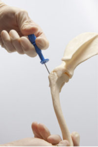
To isolate MSCs from bone marrow, a procedure referred to as a bone marrow aspiration is performed. In dogs, this procedure is typically performed on the humerus (the bone just below the shoulder) or the iliac crest (point of the hip). Dogs are placed under heavy sedation and a small patch of hair is clipped over the donor site. Local anesthetics (as one would receive at the dentist) are used to block the nerves of the skin and bone and the skin is prepared for the procedure with a surgical scrub. A needle is placed through a small skin incision (2 mm in size) into the soft, spongy bone (called cancellous bone) of the humerus or iliac crest (Figure 5). This type of spongy bone contains the largest supply of bone marrow. A small volume of bone marrow is removed with a syringe, the needle is removed, and the skin incision is closed with tissue glue. Bone marrow aspiration typically takes 2-5 minutes and, in contrast to what is reported during the procedure in humans, does not cause observable pain in dogs when performed with the use of sedation and anesthetics (pain medications). General anesthesia and surgery are not required.
Figure 5: Needle aspiration of bone marrow from the humerus (upper arm). Bone marrow MSCs can be isolated from many locations in dogs. The proximal humerus is a preferred location because of the large amount of bone marrow present and the limited soft tissues coverage of the area. Bone marrow aspiration in the dog usually takes 2-5 minutes using deep sedation and pain medications. General anesthesia and surgery are not required.
Once the bone marrow sample is isolated, it is taken to the laboratory where red blood cells are separated from the nucleated cells using a technique known as Ficoll centrifugation. The nucleated cell fraction contains marrow-derived MSCs (Mesenchymal Stem Cells), but also includes white blood cells, fibroblasts, endothelial cells, hematopoietic stem cells (HSCs), and smooth muscle cells. All of these nucleated cells are isolated, washed, counted, and plated onto sterile tissue culture dishes containing growth media. The following day, the majority of cells are washed away and a small number of cells remain tightly adhered to the tissue culture surface. These plastic-adherent, spindle shaped cells are MSCs. The MSC population is allowed to expand and once sufficient numbers of cells are present, the MSCs are lifted from the tissue culture plastic for characterization, long-term storage, use in cell culture experiments, or use in clinical patients.
Q: How are stem cells (MSCs) isolated from fat or other tissues?
Stem cells can also be isolated from fat and other tissues. To obtain MSCs from fat or other tissue, dogs are placed under general anesthesia and the skin is clipped and prepared for surgery. A skin incision is made and a small sample of fat or other tissue is removed. The fat sample is placed in a sterile container and taken to the laboratory. Once the tissue sample is in the lab, it is weighed, washed, and mechanically and enzymatically disrupted to separate fat cells and red blood cells from the Stromal Vascular Fraction of cells. The Stromal Vascular Fraction, or SVF, is a mixture of cells including white blood cells, fibroblasts, endothelial cells, MSCs, and smooth muscle cells. It is important to note that the SVF is commonly used as a therapeutic population of cells in human and veterinary cell therapy. In fact, most commercial vendors that supply stem cells for veterinary provide a preparation of SVF cells from fat tissue.
When the SVF is plated on tissue culture plastic as described above, a sub-population of the SVF adheres to the plastic and many of the SVF cells are washed away during the exchange of culture media. The remaining cells are fat-derived MSCs. When MSCs are isolated from fat, they are often referred to as ASCs or Ad-MSCs. Both SVF cells and ASCs can be used for cell therapy, however, their properties and regenerative abilities may be different from MSCs obtained from bone marrow and other tissues.
Q: Are stem cells (MSCs) isolated from various tissues the same?
Based on current information, it is clear that not all adult stem cells are identical. Tissue source, patient age, and presence or absence of disease appear to affect the number and differentiation capacity of stem cells isolated from adult dogs. This may be important when considering stem cell therapy for a pet with severe orthopedic disease. As shown in Figure 6, MSCs are much more readily isolated from canine fat or synovium as compared to bone marrow. However, when these same cells are compared using a functional assay of bone differentiation, synovial and marrow MSCs exhibit higher levels of ALP activity (a fundamental property of bone), when compared to fat-derived MSCs. Whether these differences are relevant in the context of clinical therapy has yet to be determined, but it appears that cells obtained from different tissues are not identical, even when obtained from the same donor.

Figure 6: Comparison of cell yield and bone forming potential for stem cells (MSCs) isolated from various tissues.
PANEL A: MSCs were isolated from the synovial membrane (joint lining), bone marrow, and subcutaneous fat from a single donor. MSC isolation was superior for synovium and adipose (fat) tissue as compared to bone marrow.
PANEL B: The same cells isolated in panel A were compared in an ALP activity assay, an assay of osteogenesis (bone differentiation). Synovium and bone marrow MSCs exhibited elevated ALP activity in response to increasing doses of the growth factor BMP-2, whereas little activity was present in adipose-derived MSCs.
Q: In the context of orthopedic disorders, what is stem cell therapy?
Stem cell therapy is the use of MSCs, ASCs, or SVF cells for the symptomatic or regenerative treatment of bone and joint disorders. Either autologous or allogenic stem cell therapy can be used. Autologous therapy refers to harvesting cells from one dog and re-administering those cells to the same dog. Allogenic therapy refers to harvesting cells from one dog and administering those cells to another dog, similar to a blood transfusion.
Stem cells potentially treat orthopedic disorders through a number of mechanisms, two of which are tissue regeneration and production of anti-inflammatory proteins or growth factors (Figure 7). With a tissue regeneration strategy, cells are induced toward bone or cartilage cells in the lab in conjunction with three-dimensional tissue lattices to form tissue engineering constructs for bone and joint defects. With the anti-inflammatory protein and growth factor strategy, cells are injected into diseased joints and tissues to produce proteins and other substances that suppress inflammation and improve the body’s own healing mechanisms. These mechanisms are not independent, as cross-talk between the two strategies exists. For example, cells delivered in tissue regeneration constructs likely produce specific growth factors and anti-inflammatory agents to improve healing, while cells delivered for their anti-inflammatory effects may undergo some degree of differentiation based on the specific cues provided by the treated joint or tissue.

The majority of ‘stem cell therapy’ currently being performed for canine orthopedic disorders relies on the anti-inflammatory protein and growth factor mechanism. Treatment of osteoarthritis secondary to hip dysplasia, elbow dysplasia, and cranial cruciate ligament rupture are common examples. In contrast, orthopedic disorders such as osteochondrosis (a disorder of joint cartilage) and non-union fractures (broken bones that fail to heal) are more likely candidates for tissue regeneration approaches.